Macular Degeneration
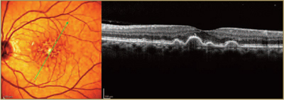
Macular degeneration is a deterioration or breakdown of the macula. The macula is a small area in the retina at the back of the eye that allows you to see fine details clearly and perform activities such as reading and driving. When the macula does not function correctly, blurriness, dark areas or distortion can affect your central vision. Macular degeneration affects your ability to see near and far and can make some activities — like threading a needle or reading — difficult or impossible.
Many older people develop macular degeneration as part of the body’s natural aging process. There are 2 types of macular problems, Dry and Wet forms. Approximately 90% of patients have the Dry form which progresses slowly. Vitamins may slow the progression in 20-25% of patients. The Wet form occurs in approximately 10% of patients and can cause bleeding and sudden loss of vision. Macular degeneration is the leading cause of severe vision loss in Caucasians over 65.
 Macular degeneration can cause different symptoms in different people. The condition may be hardly noticeable in its early stages. Sometimes only one eye loses vision while the other eye continues to see well for many years.
But when both eyes are affected, the loss of central vision may be noticed more quickly. Some common ways vision loss is detected: words on a page look blurred; a dark or empty area appears in the center of vision; straight lines look distorted.
Diabetic Retinopathy
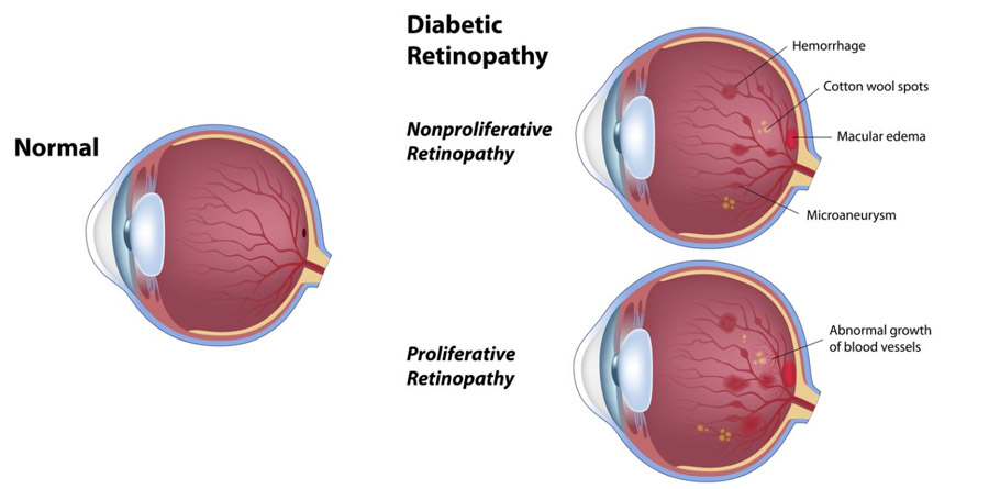
Diabetic retinopathy is a degenerative eye disease characterized by damaged blood vessels and abnormal blood vessel growth. In most cases, good management of diabetes will be enough to delay or prevent the onset of diabetic retinopathy. Yearly eye exams are recommended for all diabetic patients. These yearly eye exams will allow early treatment of damaged blood vessels and help prevent significant loss of vision. Treatment includes laser photocoagulation, steroid injections, and vitrectomy.

Laser Photocoagulation
Photocoagulation is usually effective in the treatment of Diabetic Retinopathy. The progressing damage to the blood vessels in the eye can be slowed with this treatment.
Before the surgery, eye drops are used to dilate the pupil and to numb the eye. A powerful laser beam is then focused on the damaged retina. The laser beam seals the weak or leaking blood vessels, stopping their growth.
In advanced cases of proliferative diabetic retinopathy, a vitrectomy may be recommended. A vitrectomy removes the blood-filled vitreous and replaces it with a clear solution. A sophisticated microscopic instrument is used to extract blood and scar tissue along with the natural vitreous gel of the eye. This microsurgical procedure is performed in an outpatient surgical operating room.


